Stroke Awareness Month
May 14, 2021


Types of Strokes
Ischemic Stroke
Ischemic strokes make up the majority of strokes. It occurs when a blockage obstructs a blood vessel that supplies blood to the brain. The blockage is typically comprised of fatty deposits that accumulate along the walls of blood vessels, called atherosclerosis. Blood can also clot at the location of the fatty deposit, which blocks the flow of other blood cells from reaching their target. A blood clot can form in another part of the body and travel until it reaches a vessel that is too small for it to fit through. Usually the heart and other large arteries are the source of these blood clots. Atrial fibrillation (A-fib) is a common cause of this type of embolism.
Hemorrhagic Stroke
Hemorrhagic strokes are not as common as an ischemic stroke. They are caused by a ruptured blood vessel that bleeds into the brain. As the bleeding continues, it compresses the nearby brain tissue. This is what causes the brain to shut down, leading to permanent brain damage. An aneurysm, or gradual ballooning of the blood vessel, weakens the vessel wall until it cannot withstand the blood pressure any longer. When the blood vessel bursts from an aneurysm, a hemorrhagic stroke occurs.
The Effect of a Stroke on Vision
The location of where the stroke occurs determines if and how vision is affected.
The optic nerve is the cable that connects the eye back to the occipital lobe of the brain, which is where vision is processed. If a stroke occurs anywhere near this pathway, a visual field defect (or loss of vision in a particular location) can occur. Due to the pathway of the visual system, a stroke of the right side of the brain actually affects the left visual field and vice versa.
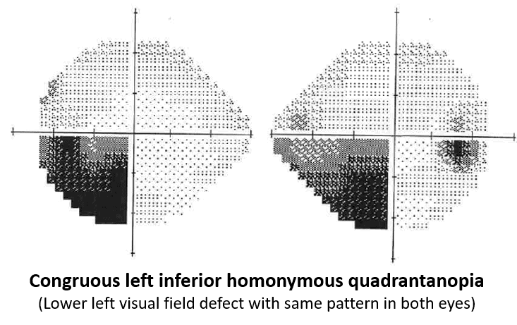
A visual field test plots a map of a person’s peripheral and central vision. The location of a stroke or mass can be identified based on the results of this test. Thankfully, MRI technology can give a much more precise location, however, the visual field is a useful tool if a patient comes to their exam with vision loss without knowing they had a stroke. Below is an example of a visual field map. The dark area in the lower left quadrant of both eyes is the location of the visual field loss.
Stroke Within the Eye
The blood vessels in the retina are some of the smallest in the whole body. This makes it relatively easy for potential blockages to occur. If the retina and optic nerve don’t receive the necessary amount of blood, containing oxygen and nutrients, the nerve tissue begins to die and permanently lose its function. There are no pain receptors in the retina, making the condition painless.
Uncontrolled hypertension, carotid artery disease, diabetes, and heart disease are major risk factors for experiencing an eye stroke. The following conditions are the main types of stroke that can occur in the eye.
Central Retinal Artery Occlusion (CRAO)
This condition occurs when the main (or “central”) artery that supplies the retina is blocked. A clot or embolus from the carotid artery or heart are the most common culprits. It only takes 90 minutes of impaired blood flow to cause irreversible vision loss. There is no treatment for a CRAO.
Branch Retinal Artery Occlusion (BRAO)
Rather than the blockage occurring in the central retinal artery, it occurs in a smaller “branch” retinal artery that supplies blood to a smaller portion of the retina.
Central Retinal Vein Occlusion (CRVO)
On the contrary to a CRAO, a CRVO involves the main retinal vein being occluded. This can also lead to bleeding in the retina, leaky blood vessels growth, and macular swelling. Eye injections are used to treat the swelling, however, it will not help any areas of the retina that have already been deprived of blood flow for an extended time.
Branch Retinal Vein Occlusion (BRVO)
In a BRVO, the blockage occurs in a smaller branch retinal vein. Bleeding and ischemia only occur in the segment of the retina that is supplied by the specific blocked branch retinal vein.
When a stroke occurs in the eye, the primary symptom will be decreased vision in the affected eye. It is important to visit your eye doctor when any sudden vision loss occurs, so the cause can be determined. Ocular strokes are typically an indication that the individual’s overall health is at risk. If this type of damage is happening in the eyes, it is certainly happening in other parts of the body too. Delayed treatment of the underlying cause can lead to an increased risk for a major stroke of the brain.
If you or someone else is experiencing symptoms of a stroke, follow the “F.A.S.T.” warning signs.

Face Drooping: Does the face droop to one side or appear uneven?
Arm Weakness: Does one arm tingle or drift downwards when both arms are lifted up?
Speech: Is speech slurred or abnormal?
Time to Call 9-1-1: If any of these signs are present, call 9-1-1 immediately. FAST treatment is important!
Resources:
https://www.stroke.org/en/about-the-american-stroke-association/american-stroke-month