The second blog of our Dry Eye Series focuses on diagnosing dry eye syndrome. Before even looking at the eyes, listening to the patient’s symptoms can direct the doctor towards the diagnosis. Although the symptoms of dry eyes can be different for each person, there are classic signs that aid in the diagnosis of the condition. The key is correlating the patient’s history and symptoms with signs present during the exam.
Common symptoms of dry eye syndrome include burning, foreign body sensation (feeling like something is in the eyes), grittiness, light sensitivity, itching, and fluctuating vision. Blurry vision that improves upon blinking can also be a symptom of dryness. Ironically, watering of the eyes can signify dryness. The eyes send a signal to the brain that more tears are needed. In response, the brain sends a rush of reflex tears that are poor quality. Rather than sticking on the eyes, these tears run down the cheek, hence, the watering.
The following methods are used in diagnosing dry eye syndrome.
Comprehensive Eye Exam
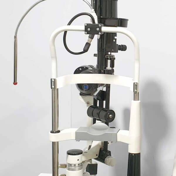
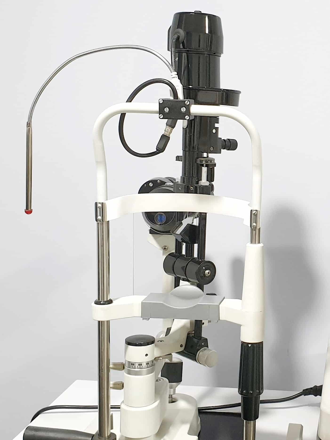
During a comprehensive eye exam, dryness can be detected during the slit lamp examination. The slit lamp is a light that uses magnifiers to give the doctor a much closer view of the eye. This allows for assessing the eyelid anatomy and looking for blink abnormalities. Some people don’t blink fully, while others may sleep with their eyes partially open. This can lead to dryness in the lower part of the cornea that is exposed all night. Dryness can affect the results of the refraction, which is the determination of the glasses prescription (“Which is better? 1 or 2?”). Therefore, it is better to seek treatment for dry eye syndrome prior to having a routine exam.
Tear Break-Up Time (TBUT)
A yellow dye, called fluorescein, is inserted into the eyes and examined using a cobalt blue light on the slit lamp. The dye gives the clear tear film a yellow color, which makes it easier to assess. In order to quantify the TBUT, the doctor counts how long it takes for the dye to dissipate immediately after a blink. This is equivalent to the amount of time it takes for the tears to evaporate into the air. If a dry spot begins to appear before 10 seconds have elapsed, the individual has dry eyes. An abnormal TBUT can indicate evaporative dry eye syndrome.
Fluorescein Staining
After assessing the TBUT, the same fluorescein dye can be used to observe the condition of the corneal surface. This test is also performed with the slit lamp, so it is commonly paired with measuring the TBUT. The fluorescein highlights patterns of dryness in the outermost layer of the cornea, called the epithelium. The location of corneal abnormalities can help determine the cause or source of dryness. For example, if the signs of dryness are in the lower one-third of the cornea, it could be due to incomplete blinking, exposure while sleeping, or a decreased blink rate.
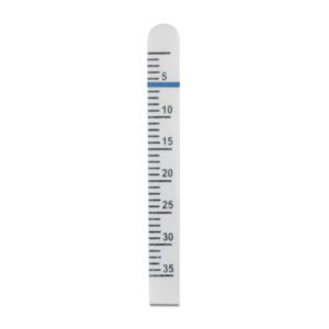
Schirmer’s Test
This test measures the volume of tears on the front surface of the eyes. Abnormal results suggest dry eye derived from decreased tear production. A thin strip of special paper with ruler-like markings is placed in the pocket of the lower eyelid and hangs down over the lower eyelid onto the upper cheek. An anesthetic eye drop can be used to decrease the amount of “reflex” tearing caused from the irritation of the paper in the eye. This tends to be a more reliable method of testing. After 5 minutes, the distance that the tears migrated down the paper is measured with the millimeter markings. If the tears wet more than 10 mm of the strip, the results are normal. Anything between 5 to 10 mm is considered a sign of mild dry eyes and less than 5 mm is indicative of severe dry eye syndrome.
Phenol Red Thread Test
Similar to Schirmer’s Test, the Phenol Red Thread Test also measures the volume of tears. A string containing phenol red dye is placed in the pocket of the lower eyelid, hanging over the eyelid like the Schirmer’s strip. When the phenol red dye comes in contact with the tears, the string turns red. After 15 seconds, the red part of the thread is then measured. Anything greater than 20 mm signifies normal tear production, while lesser amounts imply dryness. This test is not commonly used since there are other, more reliable, tests to diagnose dry eyes.
Tear Osmolarity
Measuring tear osmolarity is a newer technology with mixed reviews in terms of its importance in the role of diagnosing dry eyes. A handheld device is used to collect tears from the corner of the eye. Within seconds, the machine analyzes the tear osmolarity or the amount of particles in the tears. People with chronic dry eyes can have an imbalance of water and tear composition (salts, minerals, inflammatory cells, etc.). A higher tear osmolarity leads to inflammation, which causes dry eyes and continues the cycle of maintaining a higher tear osmolarity.
If you think you might be suffering from dry eye syndrome, call our office to schedule a dry eye evaluation. The next blog in our Dry Eye Series will cover the various treatments for dry eyes.