Optical coherence tomography (OCT) is a painless, non-invasive tool that is commonly used to diagnose and monitor changes in those with retinal conditions. Nothing touches the eyes, and there is no puff of air. Using light waves to obtain cross-sectional scans of the retina, an OCT can quickly capture 25+ scans in a matter of seconds. The horizontal and vertical cross-sections of the retina provide detailed measurements of individual retinal layers, guiding the physician to a faster and accurate diagnosis. Three-dimensional mapping of the retina and optic nerve is also a beneficial feature when viewing an elevated or depressed location. If there is something blocking the view of the retina, such as a dense cataract, the light waves will not be able to penetrate to the back of the eye, therefore, causing a poor quality scan.
iWellness Exam
If you have been to our office, you are aware of the iWellness exam that we recommend to our patients. Using OCT technology, the iWellness is a very useful screening tool for early detection of common conditions, such as macular degeneration, diabetic retinopathy, and glaucoma. Results are compared to a normative database to determine if the measurements are normal, borderline, or outside of normal limits. Scans are also compared to those of previous years to determine if there is a trend or gradual change over time. If something is abnormal, there are more detailed OCT scans that can be completed for additional data. Early detection can lead to faster treatment, which in most cases, ultimately decreases the amount of retinal or optic nerve damage and permanent vision loss.
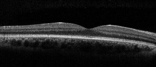
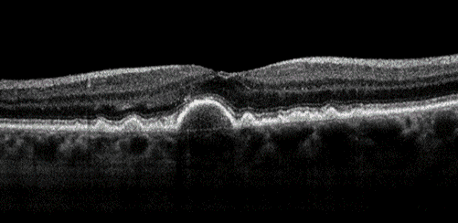
This is an iWellness scan of a healthy macula. The dip in the center of the picture is the fovea, which is responsible for precise central vision. Individual cell layers of the retina are seen as the horizontal stripes. Blood vessels are present below the retina.
Optic Nerve Scans
Optic nerve OCT is the standard of care for monitoring any progression related to glaucoma. It can also be useful in tracking changes from multiple sclerosis affecting the optic nerve. Thinning of the retinal nerve fiber layer (RNFL) is one of the key aspects in diagnosing glaucoma. In order to have vision, light hits the retina, and the signal travels through the nerve fibers to the optic nerve, which sends the signal to the back part of the brain responsible for vision. The brain interprets and processes the picture signal, giving vision in return. If the RNFL becomes damaged or thinned, the corresponding area of the visual field is compromised. OCT sensitivity picks up any changes to the optic nerve fibers before vision loss is detected on a visual field test. Glaucoma is a quiet disease with no apparent symptoms until the permanent vision loss has already become severe.
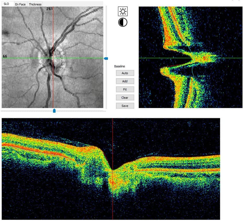
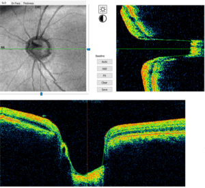
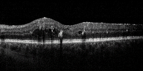
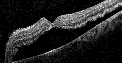
This is an optic nerve OCT of a healthy optic nerve.Compared to the normal optic nerve OCT, this patient has glaucoma with large/deep optic nerve cupping.
Retina Scans
In cases where a retinal condition is being monitored, a specific retina scan can provide more details compared to the iWellness screening tool. Macular degeneration, diabetic retinopathy, central serous chorioretinopathy, and macular holes are all conditions that warrant the use of a retina (or macula) OCT scan. The machine can also be used to scan other areas of the retina besides the macula. This can be helpful in determining whether there is fluid or elevation under a choroidal nevus (similar to a freckle below the retina) or pigmented lesion.
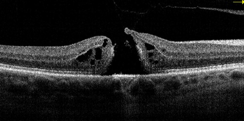
This is a retina OCT depicting dry macular degeneration.The large gap and adjacent swelling is indicative of a macular hole.
The black pockets are areas of macular edema (swelling), and the white dots are exudates (leakage of lipids) in a patient with diabetic retinopathy.This is a retinal detachment that has specifically affected the macula. Even after retinal surgery, this patient can only see the “Big E” on the chart.
An OCT scan is not a replacement for a comprehensive dilated eye exam. iWellness goes above and beyond the usual exam to help detect certain eye diseases sooner. Have you gotten the iWellness OCT yet? Call our office (717-652-7710) to schedule your next eye exam!